Home | A New Approach | Specific Up C Techniques | Blair Upper Cervical
BLAIR UPPER CERVICAL - DR. WILLIAM G. BLAIR

 The
Blair Upper Cervical approach is unique in the UpC chiropractic
profession; in that today it is still the only NON-orthogonal based
precision Spinographic (spinal radiographs or X-Rays) and adjustment
technique. In geometry ‘orthogonal’ means ‘involving
right angles’, and orthogonal based UpC techniques require
that a line drawn through the middle of the skull be perpendicular
to the plane of the atlas vertebra, which should be parallel to
the ground. Further orthogonal approaches may require the atlas
(in the vertex X-ray view) to again be at right angles to a line
drawn through the centre of the skull, although there are now considerations
being made with regards to condylar offset. This orthogonal, symmetrical
thinking is really the main departure of Dr. Blair’s approach
to that of orthogonality as Dr. Blair argues, and I think rightly
so, that the human spine is more asymmetrical rather than symmetrical.
Nevertheless, all specific UpC techniques are having a good amount
of success. The Blair technique still addresses the top two cervical
vertebrae being the atlas and axis for these are the only two which
do not have intervertebral discs, like the rest of the human spine;
are the two most freely moveable vertebrae; are the ones most commonly
misaligned and the easiest to be misaligned. Misalignments in other
spinal vertebrae, in my opinion, require far more force to occur
and are usually as a result of significant trauma. They are usually
secondary to an upper cervical subluxation. The
Blair Upper Cervical approach is unique in the UpC chiropractic
profession; in that today it is still the only NON-orthogonal based
precision Spinographic (spinal radiographs or X-Rays) and adjustment
technique. In geometry ‘orthogonal’ means ‘involving
right angles’, and orthogonal based UpC techniques require
that a line drawn through the middle of the skull be perpendicular
to the plane of the atlas vertebra, which should be parallel to
the ground. Further orthogonal approaches may require the atlas
(in the vertex X-ray view) to again be at right angles to a line
drawn through the centre of the skull, although there are now considerations
being made with regards to condylar offset. This orthogonal, symmetrical
thinking is really the main departure of Dr. Blair’s approach
to that of orthogonality as Dr. Blair argues, and I think rightly
so, that the human spine is more asymmetrical rather than symmetrical.
Nevertheless, all specific UpC techniques are having a good amount
of success. The Blair technique still addresses the top two cervical
vertebrae being the atlas and axis for these are the only two which
do not have intervertebral discs, like the rest of the human spine;
are the two most freely moveable vertebrae; are the ones most commonly
misaligned and the easiest to be misaligned. Misalignments in other
spinal vertebrae, in my opinion, require far more force to occur
and are usually as a result of significant trauma. They are usually
secondary to an upper cervical subluxation.
MALFORMATION INFLUENCE ON SPINOGRAPHIC
INTERPRETATION
Dr. Blair developed
his approach to UpC, he says in his paper “A Synopsis
of the Blair Upper Cervical Spinographic Research, November
1964, “after studying hundreds of cranio-atlanto-axial
Spinographs, … and a specimen from B.J. Palmer’s
Osteological Collection”. He discusses in his paper
his view that whilst “many systems of Cervical Spinographic
Analysis” are “theoretically, mechanically correct
… in many instances opposite conclusions are reached”
and an individual’s malformations may be being used
to formulate adjustment vectors, which might result in an
atlas being adjusted without the need for an adjustment. He
found in his research that, there was a frequency of malformations
revealing an anterior condyle occurring in 79% of all cases;
foramen magnum malformation (with the foramen magnum anterior
(A) to posterior (P) plane not parallel with the A to P sagittal
plane of the skull) occurring in 77% of all cases; a short
occipital condyle in comparison with the orbital floor in
77% of all cases; short in comparison with the base line of
the skull in 64% of cases; short in comparison with the vertical
line of the skull in 66% of the cases; transverse measurement
of the right condyle being less than of the left condyle,
in 83% of the cases. In other words “such malformations
are the rule rather than the exception” and “in
reality, one of our greatest constants is the presence of
non-symmetry in the bilateral formation of these structures.”
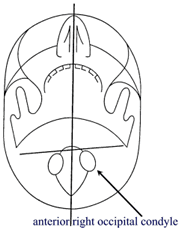
Figure 1: Offset occipital condyle
|
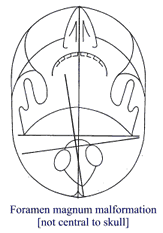
Figure 2: Offset foramen magnum |
|
OVERCOMING HAZARDS OF MALFORMATION
Dr. Blair outlines the approach to overcome possible
mistakes from misinterpreting malformations by “establishing
the Normal for each individual case” … “the normal
position of Atlas and Axis in accordance with the malformations
of each individual”. Then it is necessary to “observe
and measure the occipito (skull)-atlanto (atlas)-axial (axis) structures
to detect deviations from normal relationships.” To do this
Dr. Blair advocates the use of stereoscopic X-Ray views, which adds
a further complication since these views are very difficult to analyse.
He shifted his focus to the neurocanal, that being the canal, which
the spinal cord passes through as it makes its way from the brain
to the base of the spine. He says “Any encroachment of osseous
structure (vertebrae) into the neural canal, due to abnormal relationship
of the vertebral rings, contains the potentiality of neurological
obstruction…” and “any deviation in the continuity
of the vertebral rings can usually be detected on a stereoscopic
A to P (open mouth) cervical view and/or on the lateral cervical
flat or stereo.”
Further investigations indicate “one outstanding
feature of articulations”, “when two bones join together
to form an articulation, their two articular surfaces exactly match”
and “if any misalignment takes place it must do so at their
articulation” and further “this misalignment revealed
at an articulation gives Spinographic Science undeniable
proof of vertebral misalignment.”
MECHANICAL PRINCIPLE OF ATLAS MISALIGNMENT
The three characteristics of articular surface
of the occipital condyles are: 1. Slope; 2. Convexity; 3. Convergence.
The amount of atlas misalignment and any rotational component is
absolutely limited by the “width of the condyle-lateral mass
articular space; any further rotational movement would require a
turning of the lateral mass on the convexity of the condyle”
and “the only exception to this mechanical limitation of atlas
rotation would be condyles possessing little or no convexity”
and “we have yet to see only two cases which we feel could
have this potentiality.” In fact, if one really thinks about
the anatomy and movement as described by Dr. Blair then greater
rotation past the osseous lock of the structures and past the strength
of holding ligaments must result in dislocation of the occiput off
atlas or atlas lateral mass dislocation off axis, a situation, which
is obviously potentially fatal or neurologically damaging to a person.
The X-Ray view, Figure 3 and Figure 4 are provided
courtesy of Dr. Michael Burcon, D.C. – Blair Upper Cervical
Chiropractor- see website www.burconchiropractic.com
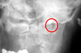 Figure
3: Stereoscopic X-Ray Figure
3: Stereoscopic X-Ray
As mentioned, the Blair technique utilises a stereoscopic X-Ray
approach shown opposite, inside the red circle. This shows the overlap
of the left occipital condyle on the left atlas condylar surface.
These articulations should line up and there should be no over or
underlap of the occipital condyles on the atlas articulating facets.
This is another look at the subluxation as described in my section “Anatomy of the Atlas Subluxation”,
but in a living person. The stereoscopic approach requires much
training and understanding of anatomy to interpret and analyse the
results. In the hands of the appropriate person this technique provides
much needed further information about the articulations in the upper
cervical spine.
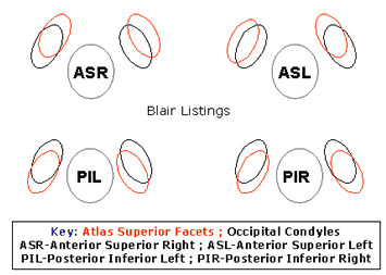 Figure
4: Blair Atlas Listings. Figure
4: Blair Atlas Listings.
The Blair chiropractor uses this and other views to prepare an atlas
listing and then finally atlas adjustment vectors. The 4 Blair Atlas
listings are described in Figure 3. The red ‘kidney shaped’
objects being the atlas superior facets and the black objects being
the occipital condyles. A head injury may result in the skull shifting
to one of these four positions, such movement dependant upon the
amplitude of the force, the direction it comes from and the anatomy
of the person sustaining the force. A consequence of the injury
can be ligament stretching, damage and/or tearing, resulting in
the person’s head remaining in a subluxated position and requiring
intervention of some kind to restore normal skull-atlas relationship
and head and neck realignment.
BLAIR CERVICAL ADJUSTMENTS
The above summary is a description of the cervical
subluxation analysis utilised in the Blair Upper Cervical approach.
The main difference from orthogonal approaches being that Blair
emphasizes the relationships of the articulating surfaces of each
vertebra. The X-Ray spinographic analysis conducted by the Blair
practitioner helps to eliminate asymmetry as a source of error in
UpC analysis.
For further analysis of the cervical subluxation
an observation of differential paraspinal dermothermograhic patterns
(DPDP) is carried out in the cervical region and functional leg
length deficiency is used. For the uninitiated DPDP utilises an
instrument that scans the cervical spine and records varying heat
patterns indicative of nerve interference. These patterns are observed
in the patient before and after cervical adjustments. The functional
leg length discrepancy comes about from the unlevelling of the pelvis
due to a misalignment at the level of the atlas as described elsewhere
on my site “Anatomy of the Atlas Subluxation”.
Together with X-Rays, and DPDP, leg length discrepancies are used
to monitor correction of the upper cervical subluxation.
The toggle adjustment is a variant on toggle-recoil,
as taught at Palmer College in the 1970’s. Blair practitioners
use the Blair Toggle-Torque (TT) adjustment approach, which was
developed from B.J. Palmer’s toggle-recoil technique. Blair
TT uses toggle without recoil but incorporates a 180-degree torque
(helical twist) force. Theoretically, "torque" is used
to decrease the superior or inferior aspect of the misalignment.
Note though that this is not torque, per se by definition as a twisting
or rotating motion about a central point. The torque may provide
extra leverage via a recto-linear force on the transverse process
of the atlas.
The patient is placed in side posture with his
or her head on a drop headpiece. The Blair TT then is delivered
by using the “pisiform lead” where the practitioner’s
pisiform bone (in the hand, next to the triquetrum and ulna bones),
remains in firm contact with the vertebral contact point throughout
the adjustment, and torque added resulting in a helical pathway.
The adjuster can alter his or her approach depending upon same side
or other side of the head, superior or inferior, anterior or posterior.
In some cases where no torque is required, the adjuster would recoil
his or her hands from the thrust.
Those who have experienced this type of adjustment
will testify that apart from a startle due to the collapse of the
headpiece, the force applied by a master of this adjustment is very
tolerable, moderately gentle and quite effective. Like the majority
of upper cervical techniques more time is spent on analysing spinographs
and other indicators than actually adjusting.
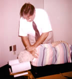 Figure
5: TOGGLE ADJUSTMENT Figure
5: TOGGLE ADJUSTMENT
The figure shows Dr. Michael Burcon (see his website - www.burconchiropractic.com)
demonstrating the Blair toggle adjustment. Michael is using the
'pisiform lead', placed upon the transverse process of his patient's
atlas. He has already determined the 'listing' of her atlas and
will make the adjustment by 'pushing' down. The headpiece (shown)
will collapse briefly and the adjustment is completed. As mentioned
previously this adjustment technique is quite tolerable, non-invasive
and involves no twisting or cracking of the neck. Following the
adjustment it is necessary for the patient to rest for 20 minutes
or so. Michael is a wonderful practitioner and has recently published
a peer-reviewed paper on Meniere's Disease; Burcon DC, Michael T.,
Upper Cervical Protocol for Ten Meniere's Patients, Journal
of Vertebral Subluxation Research - http://mccoypress.sharepoint.com/Pages/journals.aspx.I
invited Michael to Australia some years ago after corresponding
with him for almost a year on email. He was able to help a number
of my friends and neighbours; one person in particular suffering
the effects of sciatica was totally pain free within minutes of
the adjustment. I consider him a close friend. He is a graduate
of Sherman College and is a dedicated 'specific' upper cervical
chiropractor.
DOWNLOAD
PDF |
| (requires
Adobe Acobat Reader) |
blair_upper_cervical.pdf
(835kb) |
|
|
|


 The
Blair Upper Cervical approach is unique in the UpC chiropractic
profession; in that today it is still the only NON-orthogonal based
precision Spinographic (spinal radiographs or X-Rays) and adjustment
technique. In geometry ‘orthogonal’ means ‘involving
right angles’, and orthogonal based UpC techniques require
that a line drawn through the middle of the skull be perpendicular
to the plane of the atlas vertebra, which should be parallel to
the ground. Further orthogonal approaches may require the atlas
(in the vertex X-ray view) to again be at right angles to a line
drawn through the centre of the skull, although there are now considerations
being made with regards to condylar offset. This orthogonal, symmetrical
thinking is really the main departure of Dr. Blair’s approach
to that of orthogonality as Dr. Blair argues, and I think rightly
so, that the human spine is more asymmetrical rather than symmetrical.
Nevertheless, all specific UpC techniques are having a good amount
of success. The Blair technique still addresses the top two cervical
vertebrae being the atlas and axis for these are the only two which
do not have intervertebral discs, like the rest of the human spine;
are the two most freely moveable vertebrae; are the ones most commonly
misaligned and the easiest to be misaligned. Misalignments in other
spinal vertebrae, in my opinion, require far more force to occur
and are usually as a result of significant trauma. They are usually
secondary to an upper cervical subluxation.

 Figure
3: Stereoscopic X-Ray
  Figure
4: Blair Atlas Listings.
  Figure
5: TOGGLE ADJUSTMENT