 |
|
Home | A New Approach | Specific Up C Techniques | Sutter SUTTER SPECIFIC ATLAS CORRECTION - MAX SUTTER, D.C., PH.C
Max Sutter graduated from The Palmer School of Chiropractic, Davenport, Iowa in March 1925. In November 1925 he was licensed to practice Chiropractic in the State of California. His offices were opened in Huntington Park, California, in July 1926, and he was in continuous practice at that same location. Max engaged in original research from 1938 to 1940, the results of which are touched upon in the reproduction of his booklet 'The Master Key to Health' on this web page. Some of his original research provided to me by Ted and Jean Sutter, is reproduced in the downloadable file "Sutter Research". All rights are reserved. THIS I BELIEVE
SUTTER SPECIFIC ATLAS CORRECTION Although for descriptive purposes the nervous system has been divided into different parts, between the various brain and spinal cord centers and the rest of the body exists a complex interrela-tionship that binds the body into one functioning, coordinating unit. The top-most vertebra (spinal bone) of the spine is called the atlas. On either side of the large opening at the back and lower part of the head, the foramen magnum, is a bony projection called a condyle. These condyles rest upon the upper surfaces of the atlas vertebra allowing normally for a free forward and back nodding movement of the head. The second vertebra of the spine is called the axis. The head and atlas vertebra together move upon the axis in a complex pivot-ing movement when the head is turned. Although many ligaments and muscles join the head, atlas vertebra and axis vertebra move together to keep movements within nor-mal bounds and to keep the head and these vertebrae in their normal relation to each other we do not find here the same construction of bony "locks" and tough discs of fibro-cartilage that limit the move-ment of individual vertebrae from between the second and third vertebrae downward. At no other area of the spinal column do we find any degree of movement that remotely approaches that found between the head and the atlas vertebra and between the atlas and axis vertebrae. Because of the freedom of movement necessary in the region immediately above and below the atlas vertebra, and because of the construction necessary to allow for such freedom of movement, the atlas vertebra can be subluxated, that is, "locked" out of its normal articular relation sufficiently to affect vital nerve structures and thus affect the normal control of function of any organ or part of the body. Strains, variously produced, at birth, in infancy, childhood, and in adult life, affecting the atlas vertebra, may result in a sub-luxation of the atlas vertebra, and so be the initiator of an inter-ference within the nervous mechanism leading to the development of some abnormal condition of the body. Little or no local symp-toms may be present at the time the atlas vertebra is subluxated, or symptoms may fade out and disappear, and so be forgotten, and yet be the beginning of some abnormal condition that shows up in later years. So long as this subluxation of the atlas vertebra is not cor-rected, efforts to regain lost health may prove of little or no avail. The exact variation of the atlas subluxation in relation to its surrounding structures peculiar to the individual case must be ac-curately determined. This can only be done through a specialized X-ray technique and analysis. With the precise analysis of the atlas subluxation in degrees of deviation from normal articular relation the first important step in specific atlas correction has been accomplished. The specific correction of the atlas subluxation corrects the basic cause of interference within the nervous mechanism allowing the Innate Intelligence within the body to bring about a return to normal function. Specific Atlas Correction is not primarily concerned with the classification and naming of a symptom or a group of symptoms or what organ or tissues are affected, or in what manner they are affected. It realizes the great difficulty, not to say impossibility, involved in diagnosing accurately the various complex factors in-volved in each individual condition, and that the most accurate diagnosis is of little fundamental curative value unless it at the same time locates the cause of such derangement of function or structure. Specific Atlas Correction is primarily concerned with the def-inite determination of the specific subluxation of the atlas vertebra initially responsible for the development of the abnormal condition, or conditions, affecting the body, and its correction. Unless such correction has been accomplished the last word as to the ability of the Innate Intelligence within the body to bring about a natural restoration to health has not been said. Specific Atlas Correction is not concerned with palliative measures, the treatment of symptoms that may afford temporary relief, leaving the underlying disturbance as bad as or worse than before. No basically useful purpose will be served by doctoring or suppressing symptoms as long as the cause is not corrected. Only when symptoms disappear, following the correction of the cause of the condition within the body, as a result of regeneration by the Innate Intelligence within the body has anything really worth while and of lasting benefit been accomplished. Specific Atlas Correction does not concern itself with elec-tricity, radionics, lights, diet, massage, medicine, general spinal manipulation, or surgery. If you are one of the many who have failed to obtain a cor-rection of your condition through any means, Specific Atlas Cor-rection invites your serious consideration. PRACTICAL APPLICATION OF MAX SUTTER'S RESEARCH Analysis of the Atlas Misalignment
Interpretation The AP open mouth – using the anterior arch, find the center of the foramen magnum using a compass and then keeping one point at the center, compare the distance of the lateral masses of the atlas using the same structures for comparison. A significant difference will show left or right laterality of the atlas. Application Frequency
|
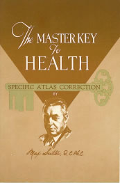
 "Your booklet (The
Master Key to Health) IS a WONDERFUL, clear, concise and
simple explanation of what and how to get sick people well.
We congratulate you on having done a GREAT job." – B.J.
Palmer
"Your booklet (The
Master Key to Health) IS a WONDERFUL, clear, concise and
simple explanation of what and how to get sick people well.
We congratulate you on having done a GREAT job." – B.J.
Palmer